Vertical Angulation in Dental Radiography: Mastering Precision to Unlock Accurate Diagnosis
Vertical Angulation in Dental Radiography: Mastering Precision to Unlock Accurate Diagnosis
In the intricate world of dental imaging, VerticalAngulationDentalRadiography stands as a pivotal technique that ensures radiographs meet the precise alignment needed to capture true anatomical relationships. This critical parameter directly influences the diagnostic value of intraoral radiographs, dictating whether a tooth’s orientation, root form, and surrounding structures appear accurately on film. Without proper vertical angulation, even the most advanced imaging systems risk producing misleading images that compromise clinical decisions—underscoring the need for meticulous attention to this often-overlooked detail.
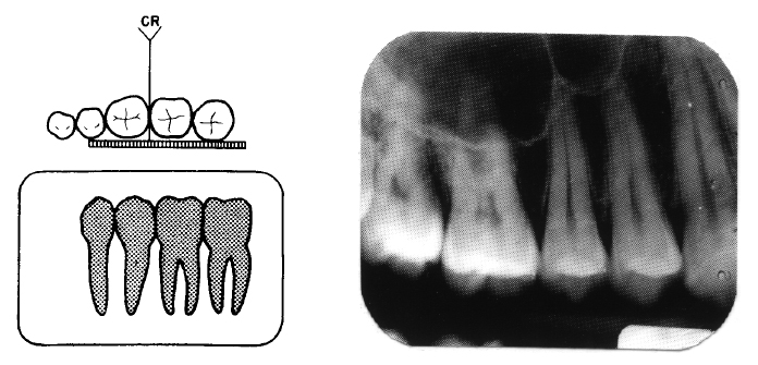
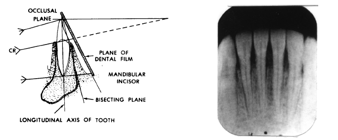
Vertical angulation refers to the angular deviation between the films or sensors and the vertical plane of the dental arch, specifically measuring how the receptor is tilted relative to the plane perpendicular to the bite axis. This alignment determines how effectively tooth positions, especially in the posterior region, are recorded—critical for detecting subtle pathologies such as periapical lesions,根分叉病变 (bifurcation defects), or impacted teeth. When the vertical axis of the receptor deviates from the ideal vertical, distortion occurs: maxillary molars may appear superimposed on posterior roots, or mandibular premolars may seem shortened or elongated, leading to misinterpretation.
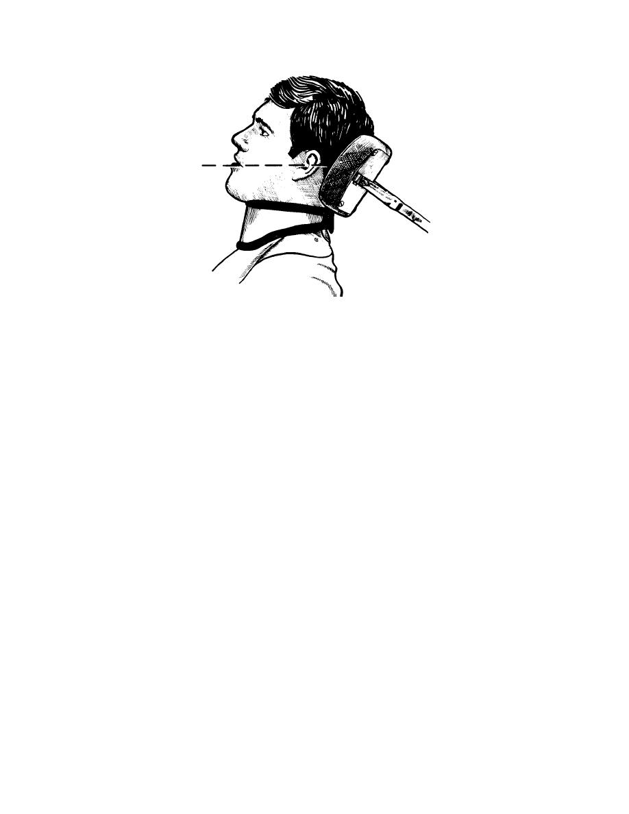
Proper vertical angulation hinges on mastering the receptor’s position relative to key anatomical landmarks. The ideal vertical alignment positions the film or digital sensor so that the upper edge aligns perfectly with the upper occlusal plane, eliminating unwanted tilt. For intraoral sensors and bitewing films, this typically means keeping the receptor parallel to the hard palate in anterior cases and level with the gingival margin in posterior regions.
In upper arch imaging, vertical angulation prevents the common “shadowing” artifact caused by upward tilt, which can obscure critical areas beneath restorations or impacted teeth.
- Use Anatomical Landmarks: Align the receptor using the pas hunched arc of the mandible or the upper labial rugae in前牙 (incisors) to maintain vertical consistency across multiple exposures.
- Employ External Angulation Guides: Many modern systems incorporate fixed angulation guides on sensor holders or sensor holders with pre-cut dovetail notches to standardize vertical positioning, minimizing operator variability.
- Leverage Digital Imaging Consistency: Digital sensors offer real-time feedback via exposure displays, allowing immediate verification of vertical alignment before sensitive exposure, reducing retakes and patient dose.
- Standardize Positioning Across Diagnoses: Establish practice guidelines for consistent vertical angles across similar anatomies—e.g., 0° tilt for O-ring or O-Vograp films on fully erupted posterior teeth—to ensure diagnostic comparability over time.
Clinical examples illustrate the profound impact of accurate vertical angulation. A study published in the Journal of Dental Radiography found that incorrect vertical alignment in posterior bitewings led to a 32% misclassification rate in detecting interproximal caries due to root overlap and artifact-induced shadowing. In contrast, images with properly calibrated vertical angles demonstrated over 98% accuracy in lesion localization.
This introduces a vertical overestimation, exaggerating root lengths and distorting adjacent anatomy. Using a tongue crib or gingival guide helps maintain consistent vertical orientation without relying solely on memory. - **Operator Fatigue and Rounding Errors Prolonged diagnostic sessions increase the risk of subtle angulation shifts over time.
Training with consistency-focused exercises—such as repeated positioning on phantom heads with marked reference lines—builds muscle memory and reduces variability between appointments, enhancing reliability across longitudinal cases. - **Failure to Adapt to Patient Anatomical Variation Children, edentulous ridges, or patients with anatomical anomalies require adjusted vertical targeting. For example, maxillary molars in higher-angulation cases (e.g., in patients with cleft alveolus or severe arch collapse) may need a −1° to −2° downward tilt to fully capture root morphology, whereas in pediatric patients, slightly upward tilting (~0° to +1°) avoids superimposition of developing teeth.
Tailoring verticle angles to individual anatomy preserves diagnostic precision without compromising image quality.
Advancements in Technology Supporting Vertical Precision
Digital radiography has revolutionized vertical angulation control by integrating software-based alignment verification. Modern systems feature built-in reference grids, automatic alignment cues, and 3D superimposition tools that overlay anatomical models onto the radiograph, highlighting misalignments in real time.Coupled with pediatric or specialty software presets, these platforms enable clinicians to achieve sub-millimeter vertical accuracy consistently, even in complex cases involving multiple quadrants or compromised patient cooperation. Ad Thousand dental practices now employ sensor holders with laser-assisted vertical alignment guides, reducing setup time by an estimated 40%. Integration with practice management software allows automatic logging of angulation parameters per patient, supporting quality assurance audits and reducing preventable diagnostic errors.
The Human Element: Skill Meets Standardization
While technology advances, the clinician’s expertise remains irreplaceable. VerticalAngulationDentalRadiography demands more than mechanical precision—it requires anatomical intuition, attention to technique, and a commitment to standardized protocols. Training programs increasingly emphasize vertical angulation mastery through simulated labs using phantom heads with variable angulations, reinforcing muscle memory and visual-kinesthetic learning.Continuous education ensures practitioners evolve with emerging tools, maintaining consistency across clinical settings. Dental professionals recognizing vertical angulation’s role in diagnostic integrity contribute directly to safer, more accurate patient care. Each radiograph, properly aligned vertically, becomes more than an image—it becomes a reliable computational canvas for detection, treatment planning, and long-term oral health monitoring.
In an era where dental diagnosis hinges on subtle visual cues, VerticalAngulationDentalRadiography exemplifies how a precise technical detail transforms routine imaging into a powerful diagnostic instrument. By championing vertical alignment standards, clinicians uphold the highest standards of care, ensuring every radiograph tells a truthful, actionable story.

Related Post

The Ultimate Noodleoscope: Noodlemagazi E—Your Global Destination for Every Noodle Trivia

Keeping Life Alive: Key Frontiers in Keap1, Kynurenine Pathway, and Kinase Biology

Elegance Woven in Fabric: The Living Heritage of Balochistan Dress

Petroleum Jelly for Burns: A Safe Relief or a Risky Misconception?