Unlocking Anatomy: A Step-by-Step Guide to Sheep Eye Dissection with Labeled Structures
Unlocking Anatomy: A Step-by-Step Guide to Sheep Eye Dissection with Labeled Structures
Peering into the intricate architecture of the sheep eye through dissection unveils a living blueprint of mammalian vision—each tissue, layer, and nerve revealed with startling clarity. Using a labeled sheep eye specimen, students and professionals alike engage with one of the most authentic biological models available, transforming abstract anatomical concepts into tangible, visible understanding. This meticulous dissection not only illuminates key ocular structures but also serves as a cornerstone for training in veterinary medicine, biology education, and surgical preparation.
Sheep eye dissection with labeled tissues is a foundational practical exercise, offering unparalleled insight into vertebrate eye morphology.The procedure systematically uncovers essential anatomical components, enabling precise identification of each part through clearly marked reference labels sited directly on the specimen. This hands-on approach bridges theoretical knowledge and real-world application, essential for aspiring ophthalmologists, researchers, and anatomy instructors alike.
The Sheep Eye: A Model for Mammalian Ocular Anatomy
The sheep eye closely mirrors the structure of the human eye in scale, tissue composition, and functional organization, making it an ideal model for dissection.
Weighing between 4 to 6 kilograms and equipped with a similar anterior-posterior orientation, the ovine eye features layers and components precisely preserved for study. Its large size facilitates detailed examination without overwhelming the learner—critical for mastering delicate dissection techniques.
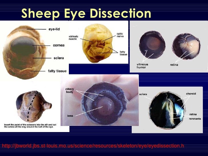
- Cornea: Transparent anterior layer, acting as the eye’s protective window.
- Iris and Ciliary Body: Pigmented structure controlling pupil size and lens shape via ciliary muscle contraction.
- Lens: Transparent avascular organ responsible for refractive focusing onto the retina.
- Retina: Light-sensitive neural layer housing photoreceptors (rods and cones).
- Optic Nerve: Bundle of nerve fibers transmitting visual signals to the brain.
- Vitreous Humor and Aqueous Humor: Fluid-filled chambers maintaining intraocular pressure and structural integrity.
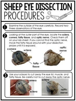
Step-by-Step Dissection Process: Achieving Precision and Clarity
Dissection begins with careful restraint and a sterile field, followed by superficial incision along the corneoscleral affront—a precise opening exposing the anterior segment.
The reflective corneal surface is lifted to reveal endothelial lining, anchoring observers to the delicate, contoured curvature central to refractive function. Higher-level structures emerge next: first, the iris—its colored pigmentation visible through a carefully removed anterior chamber—followed by the aqueous humor-filled space, often a focus of early dissection steps.
Moving deeper, observers encounter the crystalline lens suspended within the ciliary body, its elasticity pivotal for accommodation. “Separating the


Related Post

Red Oaks: The Secret Lab Where Future TV Titans Are Forged

Louisville Listcrawlers Ultimate Guide Reveals Shocking Hidden Features Hidden in U Reddit—What You’ve Been Missing

Larry Bird’s Numbers Expose a Legacy: Precision, Performance, and the DefiningIQ of a Basketball Icon

Ecuador vs. Brazil U17: The Epic Clash That Defined Under-17 Football


