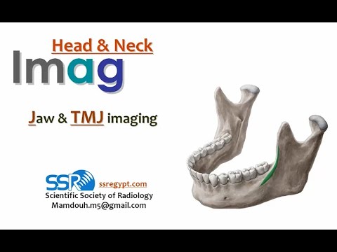
Pelvis Anatomy: Mastering X-Ray Imaging — The Radiologist’s Must-Know Guide
Pelvis Anatomy: Mastering X-Ray Imaging — The Radiologist’s Must-Know Guide
When evaluating pelvic injuries or pathologies, precise interpretation of X-ray images hinges on an in-depth understanding of pelvic anatomy and how each structure interacts radiologically. For radiologists, mastering the three-dimensional complexity of the pelvis through two-dimensional radiographic images is not just a skill—it’s a clinical imperative. The pelvis, with its intricate bony architecture and close association with vital soft tissues, presents unique imaging challenges that demand both anatomical precision and technical sensitivity.
This article distills the essential principles that enable radiologists to decode pelvic X-rays with accuracy, reliability, and diagnostic clarity.
Understanding pelvic anatomy begins with recognizing its dual role: a structural support for the spine while safeguarding pelvic organs and protecting neurovascular channels. The adult pelvis consists of three fused bones—two hip bones (each composed of ilium, ischium, and pubis)—joined anteriorly at the pubic symphysis and posteriorly by the sacrum and sacrolacunal ligaments.
This bony architecture forms a ring of 12 distinct anatomical landmarks, including the acetabulum, greater and lesser sciatic notches, and the pelvic brim, each contributing to biomechanics and imaging orientation.
Bony Landmarks and Radiographic Orientation in X-Ray Imaging
Visible on standard posteroanterior (PA) and pelvic (AP) X-rays, key pelvic landmarks serve as fixed reference points for orientation and measurement. The pelvic brim, formed by the iliac crests, defines the upper boundary, while the sacral promontory beneath provides a critical posterior anchor. The lesser and greater sciatic notches, separated by the ischial tuberosities, delineate lateral limits, useful in assessing fractures or nerve compression.The acetabulum, embedded in the ilium, is central for evaluating hip joint displacement or dislocation in trauma settings. “The pelvis is not merely a collection of bones; it’s a dynamic framework where alignment directly influences soft tissue stress and vascular integrity,” notes Dr. Elena Torres, lead radiologist at the Regional Trauma Imaging Center.
This integration of bone and function makes accurate X-ray positioning indispensable—misalignment by even a few degrees can obscure fractures or mimic pathological changes.
Positioning also impacts sensitivity: standardized views such as PA/posterior oblique (P-on-PO), AP/posterior (AP/PA), and oblique projections provide complementary views essential for full tilt assessment. Each projection reveals different aspects—PA views predominantly capture superior-posterior alignment, while oblique images bring forward anterior pelvic trauma or sacroiliac disruptions that PA projections may miss.
Key Anatomical Structures Visible on Pelvic X-Rays
Pelvic X-rays reveal not only cortical bone integrity but also the spatial relationships of adjacent structures critical to diagnosis.- **Pubic Symphysis**: A fibrocartilaginous joint between the left and right hip bones, stable in normal anatomy but prone to diastasis or widening in trauma. Radiographically identified by bilaterally symmetric posterior cartilaginous structure—widening beyond 3–4 mm raises suspicion for pubic rimming injury. - **Sacroiliac Joints (SI)**: These synarthrodial joints link the sacrum to the ilia.
In X-rays, subtle widening or asymmetric joint space narrowing suggests instability, particularly relevant in chronic lower back pain or post-traumatic injuries. - **Acetabulum**: The deep socket housing the femoral head, visible on lateral and oblique views. Fractures through the acetabular shelf or cup shape deformation indicate high-energy trauma; even hairline fractures may appear on advanced imaging.
- **Sciatic Notches and Canal**: The pelvic outlet door, where the greater


Related Post

Rob Robert Prevost Age: Decoding the Age of a Medical Science Trailblazer

Dr Doe’s Chemistry Exam: The Hidden Test That Unlocks Mastery of the Periodic Table

Mold In Toilets: The Invisible Link to Undiagnosed Diabetes Risk?

Kaitlyn Krems: The Rising Star Reshaping Social Media Influence


