Mitosis Unlocked: The Cell’s Precise Dance of Division Revealed in Stunning Cell Division Pictures
Mitosis Unlocked: The Cell’s Precise Dance of Division Revealed in Stunning Cell Division Pictures
At the core of life’s continuity lies mitosis—the meticulously choreographed process by which cells divide to ensure growth, repair, and reproduction. While often studied through textbook diagrams, the true power of mitosis emerges vividly when observed through high-resolution cell division pictures. These visuals not only capture the dynamic phases of chromosome alignment, separation, and daughter cell formation but also underscore the precision and reliability embedded in every step of human and biological development.
Mitosis, a fundamental type of cell division, transforms one cell into two genetically identical daughter cells, preserving the original genetic blueprint. This process is indispensable for multicellular organisms: it enables tissue regeneration, embryo formation, and wound healing. A single epchelle—better studied through powerful microscopic images—reveals a sequence so precise that even a single misstep risks severe consequences like cancer or developmental disorders.
Mitosis pictures, sourced from live-cell imaging and advanced microscopy, highlight each stage with striking clarity: prophase, metaphase, anaphase, and telophase, making the invisible visible and the complex comprehensible.
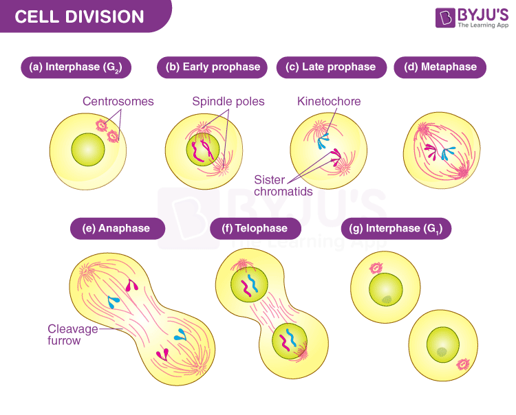
Breaking Down the Stages of Mitosis—Visualized in Detail
To understand mitosis, one must traverse its four well-defined stages, each marked by distinct structural and functional changes captured in microscopic images. - **Prophase:** Chromatin condenses into visible chromosomes, each consisting of two sister chromatids joined at the centromere.The nuclear envelope disintegrates, and the mitotic spindle begins to assemble from microtubules radiating from centrosomes. High-contrast images reveal chromosomes paired and aligned at the metaphase plate, setting the stage for accurate segregation. - **Metaphase:** Chromosomes align precisely along the equatorial plane of the cell—an elegant ballet orchestrated by spindle fibers.
These rods of protein (tubulin) pulled alternately by poles ensure parity in chromosome distribution. As described in cellular biology, “Each chromosome’s at the midpoint, poised for equal division,” a principle vividly illustrated in meticulous mitosis pictures. - **Anaphase:** Sister chromatids separate, pulled rapidly toward opposite poles by shortening kinetochore microtubules.
This dramatic movement, visible in time-lapse microscopy, demonstrates one of mitosis’s most critical feats—ensuring each daughter cell receives an identical copy. - **Telophase:** Chromatids decondense back into chromatin, nuclear membranes re-form around segregated genomes, and in some cases, the spindle disassembles. Cells begin fading from contrast as the image reflects the reassembly phase, readying for cytokinesis.
These transitions, once theories, now unfold in real time through dynamic cell division pictures, offering unprecedented insight into how life’s continuity hinges on such intricate cellular events.
How Mitosis Pictures Revolutionize Education and Research
The surge in digital microscopy and image analysis has transformed how mitosis is taught and studied. Where students once relied solely on static models and line drawings, modern learning environments incorporate interactive mitosis pictures that animate each phase.Institutions increasingly use 3D reconstructions derived from thousands of real microscopic frames, allowing learners to zoom through chromosome movement, visualize spindle dynamics, and grasp scale differences between microscale cellular events and human perception. Real time animation of mitosis confirms what biologists have known for decades: the process is both unerring and fluid. Each stage, when visualized clearly, serves as a teaching bridge—translating molecular complexity into observable phenomena.
Emerging tools now enable students to manipulate virtual cell cycles, observe mutations in chromosome


Related Post
From Cell Division’s Core: A Visual Journey Through the Phases of Mitosis—Portraits of Cell Division in Action
How Tall Is Wiz? The Unexpected Stature of a Cultural Icon

Masshealth Login

Routine of Nepal Banda: The Disciplined Path to National Cohesion and Resilience


