Inside the Mortals’ Gateway: The Anatomy of the Petrous Part of the Temporal Bone
Inside the Mortals’ Gateway: The Anatomy of the Petrous Part of the Temporal Bone
Nestled deep within the skull’s intricate framework, the petrous part of the temporal bone stands as a masterwork of evolutionary engineering—protecting delicate structures essential to hearing, balance, and sensory perception. This compact, pyramid-shaped bone segment forms the base of the temporal bone and serves as a critical anchor for vital cranial nerves and inner ear components. Far more than a passive bony shield, its complex anatomy houses sensory pathways and neural circuits that define human auditory and vestibular function.
Understanding its anatomy reveals not only physical structure but also the profound physiological roles it upholds in daily life.
Location and Structural Context
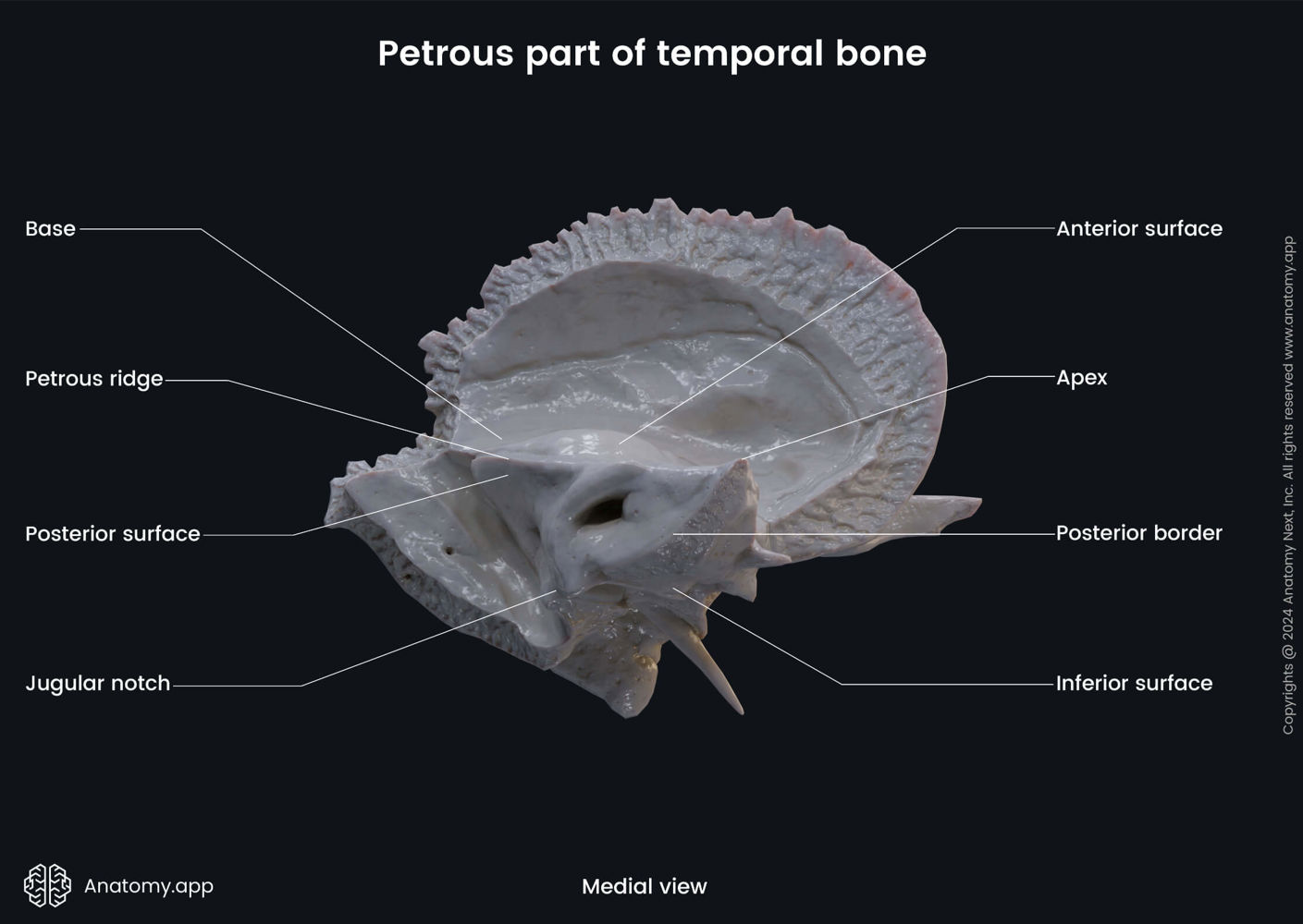
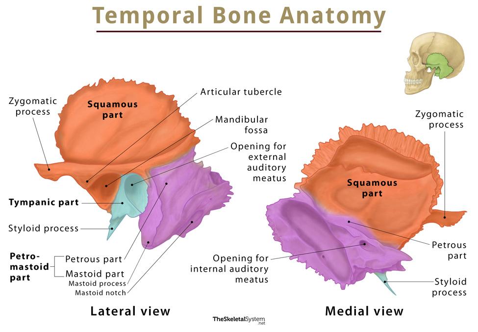
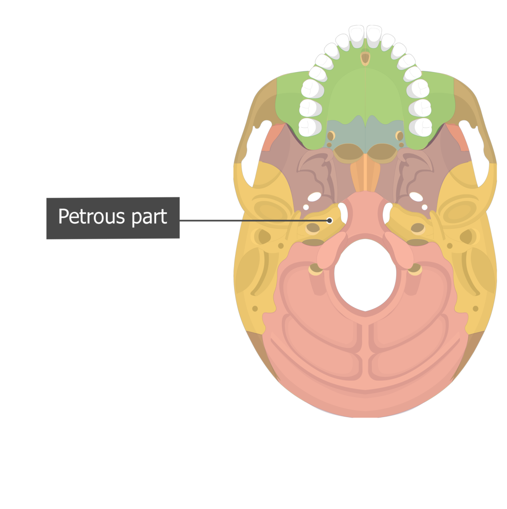
The petrous part lies at the skull base, forming the most anterior and inferior portion of the temporal bone. Transitioning from the temporal shell into the internal auditory and internal acoustic meatus, it creates a rigid framework that encases key neurovascular elements.Positioned just behind the external auditory meatus and continuous with the inner ear, this region buffers sensitive tissues from external trauma while ensuring secure passage for critical neural And vascular structures. Clinically and functionally, its strategic placement makes it a lynchpin in the transit of the facial nerve (VII), vestibulocochlear nerve (VIII), and internal acoustic vessels—all coursing through its labyrinthine passages. As neuroanatomist Dr.
Elena Moretti notes, “The petrous temporal bone is the gateway through which sensory information from the periphery enters the brainstem—its integrity is paramount for intact hearing and equilibrium.”
Microanatomy: A Labyrinth of Living Precision
The petrous bone is best understood as a microanatomical fortress composed of multiple interlocking structures. It contains two principal openings: the internal acoustic meatus, a narrow channel for the facial and vestibulocochlear nerves, and the internal auditory canal, which extends deeper toward the brainstem. These canals are lined with specialized bony trabeculae that maintain structural stability while permitting nerve passage.Encasing these pathways is a dense, mineralized bony matrix, yet remarkably perforated with small foramina—each serving a specific conduit. The internal acoustic meatus narrows sharply to just ~3 mm in width but is exactly sized to accommodate nerve fibers without compression. Venturing deeper, the internal auditory canal extends ~14 mm, decreasing in diameter to accommodate the delicate vestibulocochlear (VIII) and facial (VII) nerve roots, as well as small meningeal vessels.
Beyond these neural conduits, the petrous part contains the labyrinth—the bony labyrinth—housing the cochlea for hearing and three semicircular canals for balance. The cochlea, a coiled tube divided into vestibule, scala vestibuli, scala media, and scala tympanum, contains the organ of Corti—the sensory epithelium transducing sound vibrations into neural signals. Meanwhile, the semicircular canals detect rotational head movements, relaying motion data to the vestibular nuclei via cranial nerve VIII.
Nerve and Vessel Organization: The Neurovascular Highway
The internal acoustic meatus is a narrow corridor through which the facial (VII), vestibulocochlear (VIII), and glossopharyngeal (IX) nerves descend from the brainstem to the jugular foramen and beyond. The vestibulocochlear nerve (VIII), dedicated to auditory and balance function, divides into two branches near the petrous apex: the cochlear branch for hearing and the vestibular branch for spatial orientation. Parallel to these nerves run the internal auditory vessels—branches of the anterior and posterior cerebral arteries that supply oxygen and nutrients to the highly metabolic neuronal tissue.The nerve fibers travel within taut, lamellar sheaths created by bony trabeculae that prevent stretching or shearing. This compartmentalization—neural and vascular being carefully segregated within discrete channels—exemplifies the precision of cranial bone anatomy. With each passing nerve and vessel, the integrity of the petrous structure directly influences sensory fidelity.
Compression or damage to these conduits can result in profound deficits: facial paralysis, hearing loss, vertigo, or diplopia, underscoring the region’s critical role in neurologic and somatosensory function.
Clinical Significance and Pathological Vulnerabilities

Related Post

Top 24 News Malayalam Anchors: Who’s Shaping Kerala’s Headlines in 2024?

ITV Patrol Reveals January 7, 2023’s Shocking Steel and Systems: Key Insights That Shook the Nation

Streameast.Net: Revolutionizing Entertainment Access with Cutting-Edge Streaming Solutions

In Tears and Truth: The Emotional Depth of Love Somebody — Morgan Wallen’s Lyrics Exposed