Forms Mucous Serous and Epidermal Membranes: Nature’s First Line of Internal Defense
Forms Mucous Serous and Epidermal Membranes: Nature’s First Line of Internal Defense
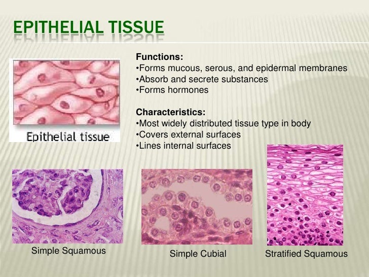
At the interface between the external world and the body’s internal systems, mucous serous and epidermal membranes form a dynamic protective barrier that defends, regulates, and sustains human health. These specialized epithelial membranes—serous, mucous, and epidermal—each serve distinct yet interconnected roles in maintaining physiological homeostasis. From lubricating airways to shielding delicate skin, their structural complexity and functional precision underpin critical bodily defenses.
Understanding their unique properties, locations, and physiological mechanisms reveals not only how the body resists infection and injury but also how clinical insights into these membranes guide medical innovation and disease management. Mucous membranes—also known as mucosae—are epithelial linings that cover internal and external surfaces exposed to the environment. Widely distributed across the respiratory, digestive, and urogenital tracts, these moist, glandular-associated tissues secrete mucus to trap pathogens, particles, and irritants.
Mucous membranes are classified by the type of fluid they produce: serous membranes secrete a thin, watery mucus rich in glycoproteins (mucins) and enzymes like lysozyme, which actively neutralize bacteria and viruses. In contrast, alveolar and gastric mucosae produce thicker, more viscous mucus containing bicarbonate ions that buffer stomach acid and protect epithelial integrity.
The serous variety thrives in enclosed spaces like the respiratory tract and conjunctiva, where rapid molecular exchange is essential, while mucous types dominate moist cavities where retention of foreign matter is critical.
Coating vital passageways from nose to bronchi and relforming linings of the stomach and intestines, mucous membranes are indispensable in maintaining sterile, functional internal environments. Unlike the rigid, keratinized surface of the epidermis, these membranes remain highly dynamic—continuously renewing and responding to environmental challenges.It is here, often overlooked, that the functional diversity of mucous membranes becomes most evident, showcasing nature’s elegant solution to balancing defense, moisture, and permeability.
Resilience in Action: The Protective Roles of Mucous Membranes
Mucous membranes serve multiple physiological functions, each tailored to the membrane’s site and secretory profile: - **Respiratory Mucous Membranes** line the nasal cavity, sinuses, and bronchi, secreting mucus that traps inhaled particles and pathogens. Ciliated epithelial cells then sweep this mucus upward—a mechanism known as mucociliary clearance—effectively sweeping away debris before it reaches deeper lung tissues. Breakdown in this system, such as in chronic bronchitis, leads to impaired clearance and persistent respiratory infections.- **Gastrointestinal Mucosae** form a critical barrier in the stomach, small, and large intestines. In the stomach, surface mucous cells secrete a bicarbonate-rich protective layer that shields the epithelial lining from aggressive gastric acid, preserving anatomical integrity despite extreme acidity. In the intestines, mucus-forming goblet cells maintain a slimy, electrically charged barrier that separates gut contents from sensitive mucosal tissue, supporting both digestion and immune surveillance.
- **Urogenital Mucous Membranes** line the urethra, bladder, and reproductive tracts, producing mucus that acts as an antimicrobial sieve and lubricant. In the female urogenital system, vaginal mucosal secretions—modulated by hormones—help maintain a balanced microbiota, reducing infection risk. - **Epi- and Metapathelial Barriers** extend beyond conventional mucosae: the epidermis itself, though primarily keratinized, functions as a physical and biochemical shield.
The stratum corneum, the outermost skin layer, consists of tightly packed, dead keratinocytes embedded in a lipid matrix that repels microbes and minimizes water loss. Beneath lies a living, regenerating epidermis where stem cells continuously renew the barrier, ensuring integrity against abrasion and infection.
Here, the epidermal membrane exemplifies passive yet indispensable protection through structural and biochemical resilience, a model of passive defense evolved for longevity.
What unites serous, mucous, and epidermal membranes is their reliance on specialized structures and secretory cells to fulfill protective roles. Mucous membranes owe their function to goblet cells and submucosal glands, which synthesize complex glycoproteins that trap pathogens and regulate surface moisture.Epidermal layers depend on keratinocytes, lipid channels, and immune sentinels to maintain impermeability and rapid repair.
These mucins form hydrated, viscoelastic layers capable of entrapment and viscous flow, crucial for expelling deleters. - **Submucosal Glands**—such as bronchial and salivary glands—secrete pre-mucosal fluids rich in bicarbonates and enzymes, which modify the gel into a stable, antimicrobial milieu. In saliva, mucins like MUC5B enable lubrication and pathogen sliding.
- **Ciliated Epithelium** lines the upper airways, synergizing with mucus to drive mucociliary currents expected to move 5–10 mm of mucus per hour in healthy lungs. - **Epidermal Kinetics** rely on keratinocyte differentiation: basal cells divide, migrate upward, and flatten into corneocytes embedded in a lipid lamellae. This process, influenced by cytokines and environmental humidity, generates a hydrophobic skin barrier, resilient to mechanical and microbial stress.
These mechanisms converge into a powerfully adaptive system—constantly remodeling in response to temperature, hydration, and immune signals. The balance of hydration, pH, and microbial flora shapes mucus quality, with disruptions—such as hypobiosis in cystic fibrosis or squamous dehydration in aging skin—compromising protective efficacy. Clinical awareness of these membranes drives targeted therapies.
For instance, hydration strategies enhance mucus rheology in chronic respiratory diseases; recombinant mucins are explored to


Related Post

Cameron Monaghan’s Wife Revealed: Everything You Need to Know About His Life Beyond the Spotlight

The Boy 2016: A Cinematic Lie That Captivated Millions and Defined a Generation

Unlock Seamless Streaming on LoJacky Roku TV: The Smarter Browser Experience Powering Modern Roku Users

Anon.Ib Maine: The Cultural Force Reshaping Maine’s Identity Through Mystery


