Areolar Connective Tissue: The Silent Architect of Cellular Support and Lead-Driven Repair
Areolar Connective Tissue: The Silent Architect of Cellular Support and Lead-Driven Repair
The areolar connective tissue, often overshadowed by more prominent connective tissues like bone or cartilage, plays a foundational role in maintaining structural integrity, enabling dynamic flexibility, and orchestrating healing processes throughout the body. Far more than passive filler, this specialized tissue serves as a dynamic interface between organs, blood vessels, nerves, and skin, ensuring seamless communication and mechanical resilience. Its unique composition of collagen, elastin, and extracellular matrix allows it to absorb stress, transmit forces, and guide tissue regeneration—functions critical to both everyday physiology and specialized repair mechanisms.
One of the defining features of areolar connective tissue is its rich network of areolar fibers—loose, interwoven bundles of collagen and elastin embedded within a hydrated matrix rich in proteoglycans and glycoproteins. This intricate lattice not only provides tensile strength to soft tissues but also facilitates nutrient diffusion and cellular migration, essential for rapid tissue response. “Areolar tissue acts as both a shock absorber and a scaffold during microtrauma,” explains Dr.
Elena Torres, a tissue biologist at the Institute of Regenerative Medicine. “It’s the body’s first responder, stabilizing injury before immune and reproductive healing systems engage.”
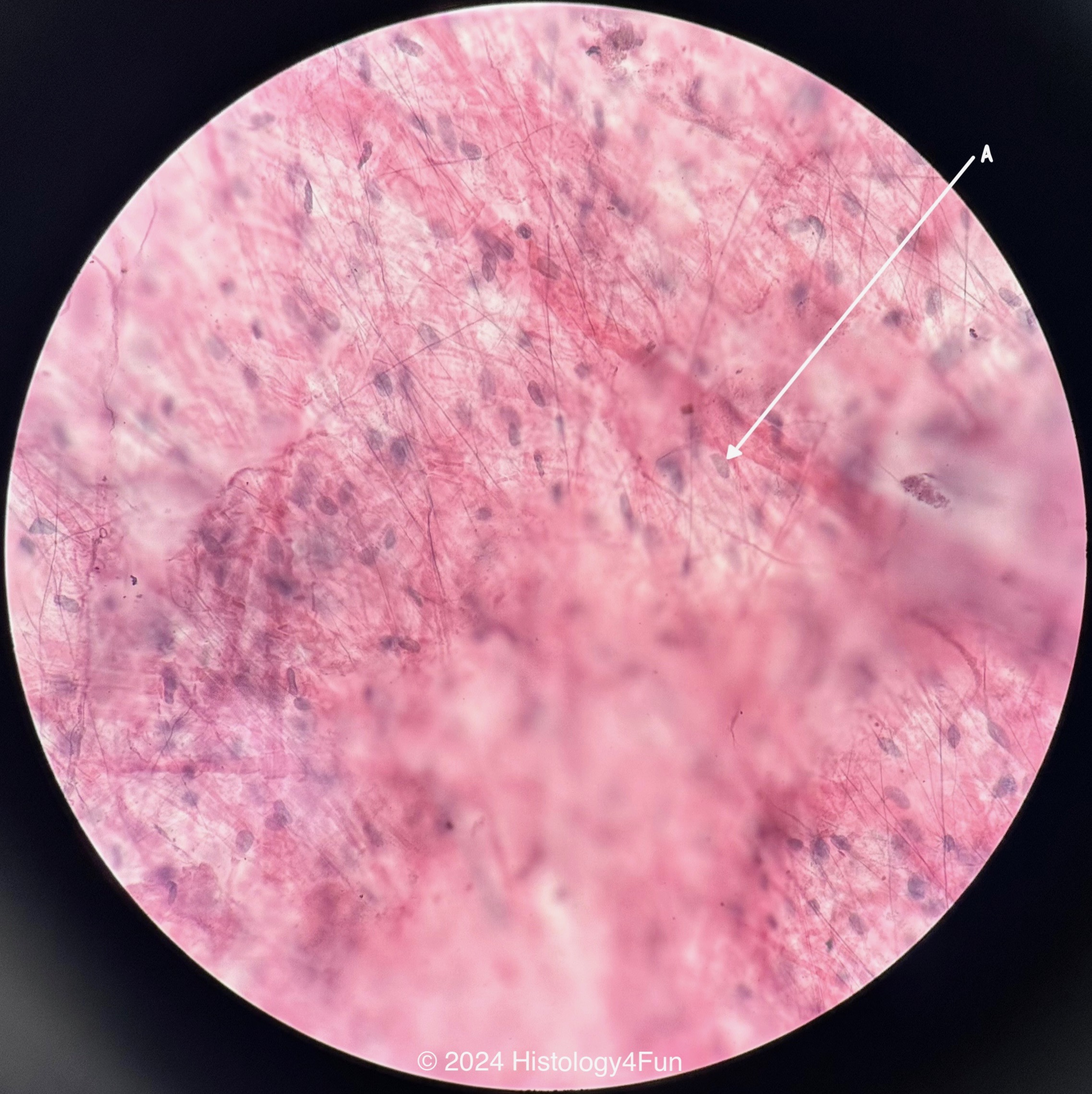
At the microscopic level, areolar connective tissue is composed of fibroblasts—the primary cellular architects—residing in oceanic lacunae and orchestrating synthesis and remodeling of the extracellular matrix (ECM). These fibroblasts dynamically adjust collagen fibril alignment in response to mechanical loading, a process known as mechanotransduction.
This adaptability allows tissues such as skin, subcutaneous fat, and organ stroma to maintain function under fluctuating biomechanical demands. The ECM’s viscoelastic properties—scanning the spectrum between rigidity and resilience—are key to its dual role in support and flexibility.
Functionally, areolar connective tissue performs several vital roles:
Mechanical Support and Force Distribution: By interconnecting muscles, blood vessels, and epithelial layers, areolar tissue disperses mechanical forces evenly, preventing localized stress fractures or tissue tearing. This is especially critical in high-motion zones such as joints and the abdominal wall.
Facilitating Cellular Communication and Migration
Adhering to the lymphatic system, areolar connective tissue serves as a conduit for immune surveillance and inflammatory signaling.
Its porous structure permits lymphocyte and macrophage infiltration during tissue injury, accelerating the transition from inflammatory to proliferative phases of healing.
Regenerative Potential and Wound Repair
During tissue injury, fibroblasts rapidly upregulate collagen production, forming a provisional matrix that guides reepithelialization and scar formation. Modern research highlights the role of growth factors—such as TGF-β and PDGF—released by resident cells, which coordinate fibroblast recruitment and ECM remodeling with remarkable precision.
Areolar tissue’s dynamic nature enables it to adapt not only to acute trauma but also to chronic mechanical stress, such as that experienced in aging skin or overuse syndromes. Studies demonstrate that prolonged strain leads to collagen cross-linking and fiber realignment, enhancing tissue stiffness and durability over time.
However, this adaptation is context-dependent—excessive stress or persistent inflammation can disrupt ECM homeostasis, predisposing to fibrosis or impaired regeneration.
Beyond structural contributions, areolar connective tissue directly influences vascular and neural function. Its tightly regulated permeability ensures optimal fluid exchange, supporting tissue hydration and metabolite transport—processes essential for maintaining metabolic equilibrium. Likewise, embedded nerve endings within the tissue allow for fine-tuned sensory feedback, contributing to proprioception and autonomic regulation of blood flow.
The Molecular Blueprint: Collagen and Elastin in Action
Collagen, constituting up to 70% of areolar connective tissue, provides tensile strength through densely packed fibrils arranged in parallel bundles.Type I collagen dominates, while Type III adds elasticity, particularly in young tissues. Elastin fibers, though less abundant, impart critical recoil properties, enabling tissues to stretch and return to base shape—a feature indispensable in skin and vascular walls. Glycosaminoglycans like hyaluronic acid anchor water molecules within the matrix, creating swelling pressures that resist compression and facilitate nutrient diffusion.
Recent advances in imaging and molecular profiling reveal how these macromolecules interact spatially and temporally during tissue repair.
Single-cell RNA sequencing of fibroblasts isolated from injured areolar tissue has identified subpopulations with specialized roles: some driving early matrix deposition, others orchestrating late-stage remodeling. This cellular heterogeneity underscores areolar connective tissue’s complexity and therapeutic importance in regenerative medicine.
Clinically, understanding areolar connective tissue function informs treatments for chronic wounds, scars, and connective tissue disorders. For instance, therapies enhancing ECM synthesis—such as platelet-rich plasma (PRP) or biomimetic scaffolds—leverage the tissue’s innate regenerative capacity.
Targeting pathological fibrosis through controlled modulation of TGF-β signaling represents a promising frontier in medicine, aiming to restore tissue balance without compromising structural support.
In summation, areolar connective tissue is a marvel of biological engineering—far more than inert connective support. Its dynamic composition, mechanosensitivity, and regenerative precision enable it to protect, adapt, and heal across a spectrum of physiological challenges. As research continues to uncover its hidden sophistication, areolar tissue emerges not just as a passive framework but as a central player in systemic health and recovery, shaping outcomes from everyday wear to complex tissue repair.


Related Post

Discover the Life of Casey Nezhoda’s Daughter: Behind the Public Face, a Story of Identity, Privacy, and Legacy

Fallen / Brand New World: Where Culture Collides with Digital Immortality

Ethan Allen Credit Card Login: Your Secure Path to Effortless Financial Access

How New York Parking Ticket Pay Simplifies City Driving & Saves Time